0%

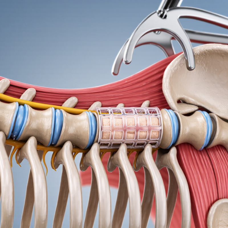
Lumbar Interbody Cages have become important tools in modern spinal fusion. They are placed between damaged vertebral bodies after the disc is removed. The cage helps restore disc height, support spinal alignment, and create space for bone growth. Imagine a small structural spacer working deep inside the lower back. Its role is precise, but never simple.

Spine surgeon Dr. Lawrence Lenke has emphasized, “Every patient is different, and every patient needs an individualized treatment plan.” That principle explains why surgeons consider more than the cage alone. They assess symptoms, bone quality, spinal balance, nerve compression, previous operations, and overall health. A well-designed cage may improve stability and help maintain alignment. It may also support indirect nerve decompression in selected patients. The surgical approach matters too. So does the patient’s recovery plan.

Still, Lumbar Interbody Cages are not automatically the best answer. Fusion can involve pain, nonunion, infection, nerve injury, or adjacent-segment problems. Results vary. The evidence is strong in some conditions, but less certain in others. That uncertainty deserves honest discussion. Surgeons must match implant design and surgical technique to the individual anatomy, not marketing language. Patients should ask why fusion is recommended, what alternatives exist, and how success will be measured. A cage is only one part of a larger biological and mechanical process. The real goal is not an impressive implant. It is durable stability, useful pain relief, and a safer return to daily movement.
Why Choose Lumbar Interbody Cages for Spinal Fusion?

What Are Lumbar Interbody Cages?

Lumbar interbody cages are small medical implants placed between two lumbar vertebrae after the damaged disc is removed. They restore disc height, support spinal alignment, and create space for bone graft material. As new bone grows around the cage, the motion between vertebrae may gradually reduce. Think of it as a carefully fitted spacer, not a simple replacement part.

Cages are commonly made from titanium, cobalt-chromium, or radiolucent polymers such as PEEK. Their surfaces may include grooves, porous structures, or coatings that encourage bone attachment. A 2023 review in the European Spine Journal reported fusion rates commonly above 85%, although results varied by surgical approach, patient health, and follow-up time. The North American Spine Society also emphasizes patient selection and imaging confirmation rather than relying on symptoms alone. That matters.

Clinical experience shows that cage height must match the patient’s anatomy. An oversized implant may increase endplate stress and cause subsidence, where the cage sinks into the bone. A 2022 systematic review in Global Spine Journal identified subsidence as a recognized complication, with rates differing substantially across techniques and definitions. Smoking, osteoporosis, and poor bone quality can further weaken stability. A cage supports fusion, but it cannot correct every biological problem. Even careful planning has limits.

How Do Lumbar Interbody Cages Support Spinal Fusion?

Lumbar interbody cages support spinal fusion by replacing a damaged disc space with a stable, bone-friendly structure. Surgeons remove painful disc material, prepare the vertebral endplates, and place bone graft inside or around the cage. The implant maintains disc height, protects the graft, and helps restore foraminal space for exiting nerves. That mechanical support matters during early healing.

The cage also shares axial load across the spine. Its textured surface and open internal space can encourage bone to grow through the implant. Correct sizing is critical. A cage that is too small may fail to restore height; excessive distraction can irritate joints or nerve roots. It is not a magic spacer.

A 2022 meta-analysis in Global Spine Journal reported fusion rates near 90% for minimally invasive transforaminal lumbar interbody fusion, although imaging methods varied between studies. The North American Spine Society’s coverage recommendations emphasize patient selection, neurological findings, instability, and failed nonsurgical care. These factors often matter more than the cage alone. The FDA also requires interbody devices to be evaluated for intended spinal-fusion use, but approval does not guarantee union in every patient.

In practical terms, successful fusion depends on cage position, endplate preparation, bone quality, graft biology, smoking status, and postoperative loading. The evidence is encouraging, yet imperfect. Radiographic fusion may not equal complete pain relief. That distinction deserves honest discussion before surgery.

Why Choose Lumbar Interbody Cages for Spinal Fusion?

How Do Lumbar Interbody Cages Support Spinal Fusion?

Lumbar interbody cages are placed between adjacent vertebral bodies to restore disc height, improve spinal alignment, maintain foraminal space, and contain bone graft. By sharing axial load with the surrounding bone and providing a stable environment for bone growth, cages can support successful interbody fusion.

The chart shows representative midpoints of fusion-rate ranges commonly reported in peer-reviewed clinical reviews: ALIF 90–100%, TLIF 90–95%, PLIF 90–95%, and LLIF 85–95%. Outcomes vary according to patient selection, surgical technique, graft material, follow-up duration, and study design.

What Conditions Can Lumbar Interbody Cages Treat?

Why Choose Lumbar Interbody Cages for Spinal Fusion?

What Conditions Can Lumbar Interbody Cages Treat?

Lumbar interbody cages may help patients with painful spinal conditions that resist non-surgical care. Common examples include degenerative disc disease, spondylolisthesis, and lumbar spinal stenosis. They can also support treatment for recurrent disc problems or instability after earlier surgery. The cage sits between two vertebrae after the damaged disc is removed. It helps restore disc height and may reduce pressure around irritated nerves. Bone growth through the cage can create a stable fusion over time.

Not every back problem requires a cage. A careful assessment usually includes symptoms, physical findings, and imaging such as MRI or X-rays. Doctors also consider walking limits, leg weakness, pain duration, and previous treatments. For example, foraminal stenosis may improve when restored disc height creates more room for a compressed nerve. Results vary. The operation is not a guaranteed cure, and fusion can place extra stress on nearby spinal levels. The choice can be difficult, even for experienced surgical teams.

Tips: Ask which condition the procedure targets and what success means for you. Discuss bone health, smoking, medications, and recovery limits before surgery. Follow movement instructions closely after the operation. Report increasing weakness, fever, wound drainage, or severe pain promptly. Clear questions often prevent unrealistic expectations.

What Benefits Do Lumbar Interbody Cages Offer?

Why Choose Lumbar Interbody Cages for Spinal Fusion?

Lumbar interbody cages support fusion between two vertebrae after a damaged disc is removed. They restore disc height and may enlarge narrowed nerve openings. This can reduce mechanical pressure around an irritated nerve. Their structure also helps transfer weight through the front of the spine, where natural loading occurs.

A 2023 World Health Organization report estimated that 619 million people experienced low back pain in 2020. The number may reach 843 million by 2050. Better surgical planning matters.

What Benefits Do Lumbar Interbody Cages Offer?

Cages can provide a stable space for bone growth. Surgeons may also use them to improve spinal alignment, especially when disc collapse causes forward bending. Clinical reviews generally report improvements in pain, function, and fusion rates, but outcomes vary by surgical approach, bone quality, smoking status, and diagnosis. Evidence is not perfectly uniform. That deserves honest discussion.

The North American Spine Society emphasizes patient selection and shared decision-making in lumbar fusion care. A cage is not a cure by itself. It works within a broader procedure involving decompression, fixation, bone grafting, and rehabilitation. In practice, patients often notice the difference through small details: standing longer, walking farther, or sleeping with fewer position changes. Still, some patients may improve without fusion, and every cage carries risks, including subsidence, nonunion, infection, or nerve injury.

What Risks and Recovery Considerations Should Patients Know?

Lumbar interbody cages can restore disc height and support fusion, but surgery is not risk-free. Nerve irritation may cause temporary leg pain, numbness, or weakness. Less common complications include infection, blood clots, dural tears, cage migration, and failed fusion. The National Surgical Quality Improvement Program reports that major complications after elective lumbar fusion generally remain below 10%, but risks increase with age, smoking, obesity, diabetes, and limited mobility.

Cage subsidence deserves attention. A 2023 systematic review in the European Spine Journal reported widely varying subsidence rates, from under 5% to more than 30%, depending on the cage position, bone quality, and imaging definition. Subsidence may cause renewed pain or reduced correction. It does not always require another operation. That distinction matters. Patients should ask how their surgeon measures it.

Recovery is rarely a straight line. Most patients walk soon after surgery, while lifting, bending, and driving restrictions may last several weeks. Bone fusion can require six to twelve months, according to guidance from professional spine societies. Physical therapy often rebuilds core strength gradually. Progress can feel frustratingly slow. Persistent fever, wound drainage, new weakness, or bladder and bowel changes require urgent medical assessment. A 2022 review in Spine found that smoking is associated with poorer fusion outcomes, so stopping before surgery is a practical step. Even careful planning cannot remove every uncertainty. The recovery plan should reflect the patient’s bone health, work demands, medications, and realistic tolerance for temporary limitations.

Why Choose Lumbar Interbody Cages for Spinal Fusion? - What Risks and Recovery Considerations Should Patients Know?

Evidence-based clinical considerations for adults undergoing lumbar interbody fusion

Clinical Dimension What Lumbar Interbody Cages Do Potential Patient Benefit Important Risks or Limitations Typical Recovery Considerations Evidence Context
Structural Support An interbody cage is placed between two lumbar vertebral bodies after removal of the damaged disc material. It helps maintain disc-space height while bone grows around or through the implant. May help restore or preserve spinal alignment and provide a stable environment for fusion. Loss of height, implant migration, cage subsidence, or failure of fusion can occur, especially when bone quality or mechanical loading is unfavorable. Patients may need temporary activity restrictions while the fusion develops. The cage provides immediate structural support, but biological fusion is gradual. The cage is not the fusion itself; successful fusion depends on bone healing, fixation, alignment, and patient factors.
Pain and Nerve Symptoms Disc removal and restoration of foraminal height may reduce mechanical disc pain and relieve pressure on affected nerve roots when nerve compression is present. Leg pain caused by nerve compression often improves more predictably than nonspecific low-back pain. Persistent or recurrent back pain may occur. Nerve irritation, numbness, weakness, or injury is uncommon but clinically important. Early soreness is expected. Nerve symptoms may improve quickly or gradually over weeks to months, depending on the duration and severity of preoperative compression. Outcome depends strongly on the underlying diagnosis, nerve health, spinal alignment, and whether pain is genuinely arising from the treated level.
Fusion Success The cage commonly contains bone graft or a bone-graft substitute and is used with additional fixation, such as pedicle screws and rods, to limit motion at the treated level. A solid fusion can reduce painful motion and help maintain correction at the operated segment. Nonunion, also called pseudarthrosis, may cause ongoing pain or hardware stress and can require revision surgery. Radiographic evidence of fusion commonly develops over approximately 6–12 months, although timing varies by patient and surgical technique. Smoking, nicotine exposure, poor bone quality, diabetes, multilevel surgery, and inadequate nutrition can reduce the likelihood of successful fusion.
Approach Selection Cages may be inserted through anterior, lateral, oblique, or posterior approaches. The selected route depends on anatomy, diagnosis, prior surgery, and surgeon assessment. Some approaches may allow better restoration of disc height or alignment while reducing direct manipulation of certain posterior muscles. Each approach has distinct complications. Anterior approaches may involve vascular, bowel, or sexual-function risks; lateral approaches may affect psoas or lumbar plexus structures; posterior approaches may involve muscle and nerve-related risks. Hospital stay and activity restrictions vary by approach and by whether additional decompression or fixation is performed. No single approach is best for every patient. Individual anatomy and the surgical indication are central to decision-making.
Infection and Wound Healing The operation involves an incision, disc-space preparation, graft placement, and usually internal fixation. Modern sterile techniques and perioperative antibiotics reduce, but do not eliminate, infection risk. Superficial or deep infection, wound drainage, delayed healing, or the need for surgical cleaning can occur. Patients should monitor for increasing redness, swelling, drainage, fever, worsening pain, or wound separation and report these promptly. Risk is influenced by diabetes control, smoking, obesity, immune status, nutrition, operative complexity, and previous spinal surgery.
Blood Clots and Medical Complications Spinal fusion is a major operation and may temporarily reduce mobility. Early mobilization, leg exercises, and individualized preventive measures can reduce postoperative complications. Deep-vein thrombosis, pulmonary embolism, urinary problems, constipation, medication reactions, and cardiopulmonary complications are possible. Walking is usually encouraged as soon as safely possible. Blood-thinning medication is prescribed selectively because it must be balanced against bleeding risk. Preventive plans depend on personal clotting risk, bleeding risk, mobility, medical history, and the surgeon’s protocol.
Adjacent-Level Degeneration Fusion removes motion at the treated segment, which may alter load distribution across neighboring spinal levels. Stabilizing an unstable or painful segment may improve function and reduce symptoms from that level. Adjacent spinal levels may degenerate over time, and some patients may later develop symptoms or require additional treatment. Long-term follow-up is important if new back pain, leg pain, numbness, or weakness develops after the initial recovery. Age-related degeneration also occurs naturally, so not every later change is caused by the fusion.
Hospital Recovery The procedure may be performed with minimally invasive or open techniques, depending on the required correction and decompression. Many patients can begin standing and walking with assistance on the day of surgery or the following day. Longer hospitalization may be necessary after multilevel surgery, significant blood loss, medical complications, or limited mobility. For uncomplicated cases, hospital stay is often about 1–3 days, but this varies substantially by procedure, health status, and local practice. Discharge generally depends on pain control, safe walking, adequate bladder function, and the ability to manage basic activities.
Return to Daily Activities The implant provides stability while soft tissues heal and the fusion matures. Gradual return to walking and routine light activities can support conditioning and independence. Bending, lifting, twisting, driving, and impact activities performed too early may increase pain or mechanical stress. Light daily activities may resume over several weeks. Return to work may range from a few weeks for sedentary duties to several months for heavy physical work. Timing should be individualized according to symptoms, neurological status, imaging, work demands, and the treating team’s instructions.
Physical Therapy Rehabilitation focuses on safe mobility, posture, walking tolerance, trunk control, and progressive strengthening. A structured program may improve function, confidence, conditioning, and body mechanics. Overly aggressive exercise can aggravate symptoms or interfere with recovery. Therapy is not a substitute for evaluation of new neurological deficits. Walking is commonly introduced early. Formal therapy may begin after the surgeon confirms adequate wound healing and provides activity clearance. Rehabilitation protocols vary; there is no universal timeline for every fusion patient.
Warning Signs After Surgery Postoperative monitoring helps identify complications before they become more serious. Prompt assessment can improve the chance of timely treatment. Urgent symptoms include new or worsening leg weakness, loss of bladder or bowel control, severe calf swelling, chest pain, shortness of breath, high fever, or worsening wound drainage. Patients should contact the surgical team immediately for emergency symptoms rather than waiting for a routine appointment. The appropriate response depends on symptom severity and local emergency-care instructions.
Factors Supporting Better Outcomes Preoperative optimization and adherence to postoperative instructions support bone healing and safe rehabilitation. Stopping nicotine, managing diabetes, maintaining adequate nutrition, treating osteoporosis, and following lifting restrictions may improve recovery conditions. Nicotine use, untreated osteoporosis, poor nutrition, uncontrolled medical conditions, and nonadherence may increase complications or nonunion risk. Patients should review medications, bone-health status, nutrition, home support, and work requirements before surgery. Individual risk assessment is more useful than relying on a single average complication rate.
Clinical note: Recovery times, complication rates, and suitability for lumbar interbody fusion vary according to diagnosis, spinal level, number of levels treated, surgical approach, bone quality, general health, and surgeon experience. This table is for general education and does not replace individualized medical advice.

FAQS

What is a lumbar interbody cage?

It is a small implant placed between two lower-spine vertebrae.

How does a lumbar interbody cage support fusion?

Bone graft is placed inside or around the cage.

Which conditions may require a lumbar interbody cage?

Possible conditions include disc degeneration, vertebral slipping, and spinal narrowing.

What benefits can these cages provide?

A cage can restore lost disc height and create more room for irritated nerves.

What materials are used in lumbar interbody cages?

Common materials include titanium, cobalt-chromium, and radiolucent polymers.

What risks should patients discuss before surgery?

Risks include cage sinking into bone, failed fusion, infection, and nerve injury.

How is a patient evaluated before receiving a cage?

Doctors usually review symptoms, physical findings, and MRI or X-ray results.

What should patients expect during recovery?

Follow movement instructions closely after surgery.

Conclusion

Lumbar Interbody Cages are medical devices used in spinal fusion surgery to replace a damaged spinal disc and help stabilize the affected area. Positioned between two vertebrae, they restore disc height, support proper spinal alignment, and create space for bone growth. Over time, new bone can grow around the cage, joining the vertebrae into a stronger, more stable segment. Depending on the patient’s condition and surgical approach, these cages may be used to treat degenerative disc disease, spinal stenosis, spondylolisthesis, recurrent disc problems, and certain spinal deformities.

Potential benefits include reduced pain caused by instability or nerve compression, improved spinal structure, and enhanced long-term support. However, patients should understand that surgery may involve risks such as infection, nerve irritation, blood clots, cage movement, or failure of the bones to fuse. Recovery varies according to overall health, surgical technique, and adherence to medical guidance. Follow-up visits, gradual activity increases, rehabilitation, and avoiding excessive strain are important for achieving the best possible outcome.


Amelia

Amelia

Amelia is a dedicated marketing professional with a strong understanding of the company’s products, market positioning, and customer needs. With a background in strategic communication, content development, and digital marketing, she transforms complex product information into clear, practical, and......